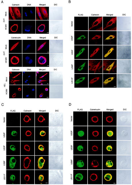
FIG. 6.
Subcellular localization of ER in HCMV-infected EVT. (A) The localization of ER in EVT and HEL was examined using anticalnexin antibody and anticalreticulin antibody. DNA was stained with Hoechst 33258. Cells were infected at an MOI of 5 and examined at 24 hpi. (B) Localization of FLAG-tagged US proteins in EVT was examined. EVT were transfected with the expression plasmids for FLAG-tagged US proteins, US2F, US3F, US6F, and US11F. The localization of FLAG-tagged proteins was detected at 24 h after transfection using anti-FLAG antibody. The localization of the ER in the same cells was examined with anticalnexin antibody. (C) Localization of FLAG-tagged US proteins and ER (calnexin) in HCMV-infected EVT was examined. EVT were transfected with the expression plasmids for FLAG-tagged US proteins and then infected with HCMV at an MOI of 5 at 24 h after transfection. The localization of FLAG-tagged proteins was detected at 24 hpi using anti-FLAG antibody. The localization of the ER in the same cells was examined with anticalnexin antibody. (D) Localization of FLAG-tagged US proteins and ER (calreticulin) in HCMV-infected EVT. EVT were transfected with the expression plasmids and then infected with HCMV as described in Fig. 6C. The localization of FLAG-tagged proteins and the ER were detected at 24 hpi using anti-FLAG antibody and anticalreticulin antibody. All cells were examined using a confocal laser scanning microscope. DIC, differential interference contrast microscopy. Scale bar, 10 mm.

