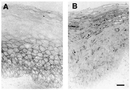
FIG. 2.
E-cadherin expression is reduced and cellular localization is altered in HPV16-infected tissue. (A) Normal epidermis showed a typical staining pattern after E-cadherin labeling, with a positive signal apparent on the cell surface of basal and suprabasal KC. (B) HPV16-infected epidermis showed punctate intracellular staining in the superficial layers of the epidermis. Bar, 10 μm.

