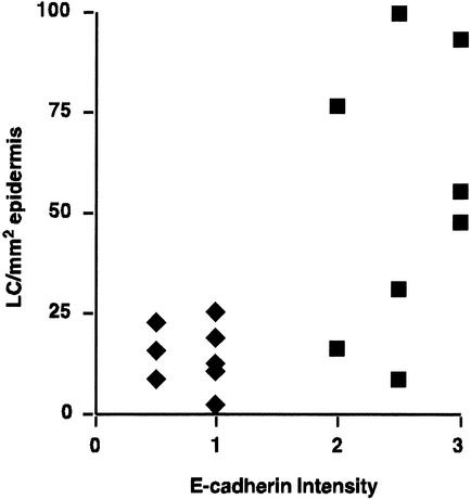
FIG. 3.
There is a direct correlation between E-cadherin expression and LC density in the skin. E-cadherin staining patterns in HPV-infected (♦) and normal tissues (▪) were graded on a semiquantitative scale (loss of staining = 0; weak and fragmented distribution = 1; heterogeneous distribution = 2; strong and normal distribution = 3) and were plotted against LC number. E-cadherin staining was low or absent in HPV16-infected tissues, and there was a low density of LC. E-cadherin staining was stronger and LC density was generally higher in normal tissues. There was a direct correlation between the level of E-cadherin and LC density in both normal and infected tissue (P = 0.01).

