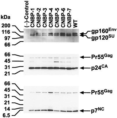
FIG. 1.
Immunoblot analysis of NC mutant and wild-type HIV-1. Proteins were fractionated by sodium dodecyl sulfate-polyacrylamide gel electrophoresis and transferred to an Immobilon-P (Millipore) membrane. A total of 6.1 × 10⁶ cpm of RT activity/ml for each sample was loaded onto the gel. The membrane was incubated sequentially with α-p7NC (bottom panel), α-p24CA (middle panel), and α-gp120SU (top panel). Bands were detected by chemiluminescence as described previously (25, 27). Molecular weight markers are indicated on the left, the virus analyzed is designated at the top, and the pertinent proteins detected by the various antibodies are specified on the right.

