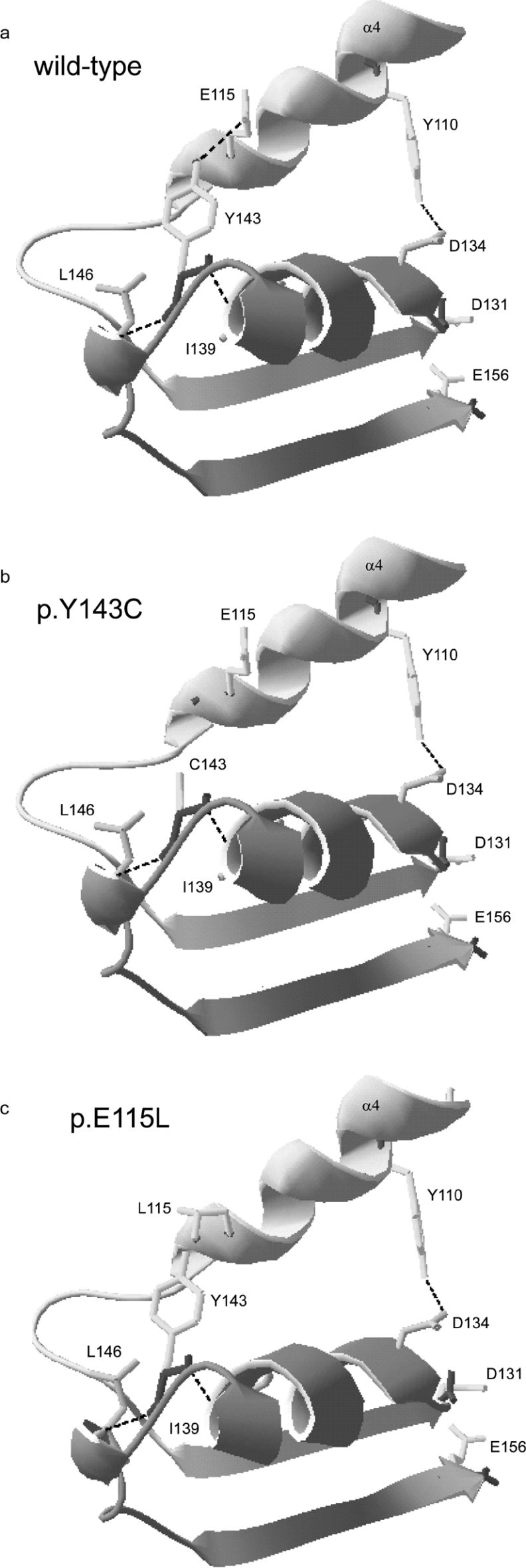
Figure 7. Ribbon diagrams of the region containing Tyr143 in human AdoHcyase.
The enlargements show the region containing residues Tyr106 to Glu156 of human AdoHcyase. Mutated amino acids or residues involved in enzymatic activity and hydrogen-bonding are represented with side chains. Existing hydrogen bonds are shown as broken lines. (a) Wild-type; (b) p.Y143C is lacking hydrogen bond between Tyr143 and Glu115; (c) p.E115L is lacking hydrogen bond between Tyr143 and Leu115.

