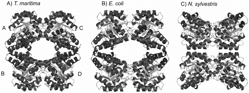
Figure 1. X-ray crystal structures of DHDPS from (A) T. maritima, (B) E. coli and (C) N. sylvestris.
Each enzyme is a homotetramer composed of two tight-dimer units (A–B and C–D), but the arrangement of the two dimeric units is different. The structures in (B) and (C) were drawn using the co-ordinates described in [38] and [17] respectively. The structures were drawn using Pymol (http://www.pymol.org).

