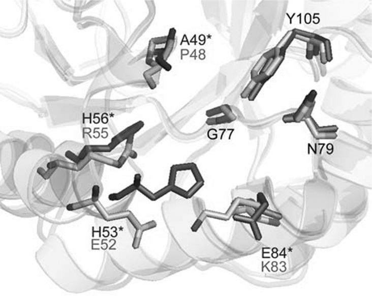
Figure 5. Overlay of the (S)-lysine binding site of E. coli (black) and T. maritima (grey) DHDPS structures.
Numbering is shown for the T. maritima enzyme, except where the residues are not conserved, in which case the E. coli numbering and residue are indicated by an asterisk. This Figure was drawn using Pymol (http://www.pymol.org).

