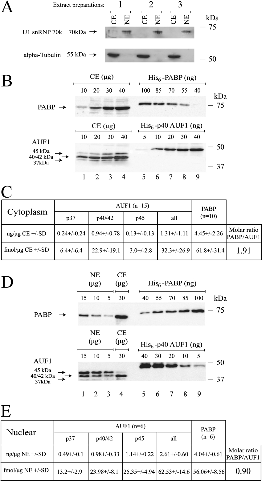
Figure 7. AUF1 is an abundant protein in HeLa cytoplasm and nucleus.
(A) Cytoplasmic extracts (CE) (20 μg) and 5 μg of nuclear extracts (NE) from three distinct preparations were resolved on a 10% polyacrylamide gel and analysed by Western blotting using either an anti-α-tubulin or anti-U1 snRNP70k antibody as indicated. (B) Various amounts of HeLa cytoplasmic proteins (CE, lanes 1–4, indicated in μg), His6–PABP (lanes 5–9, top panel, indicated in ng) or His6–p40AUF1 (lanes 5–9, bottom panel, indicated in ng) were resolved on an 8% polyacrylamide gel and analysed by Western blotting using either an anti-PABP (upper panel) or anti-AUF1 (bottom panel) antibody. (C) The Table summarizes the results from independent experiments (number as indicated) using the three distinct HeLa cytoplasmic extracts shown in (A). Results are expressed in either ng or fmol of corresponding protein per μg of HeLa cytoplasmic extract±S.D. (D) Various amounts of HeLa nuclear proteins (NE, lanes 1–3, indicated in μg) and cytoplasmic proteins (CE, lane 4, indicated in μg), His6–PABP (lanes 5–9, upper panel, indicated in ng) or His6–p40AUF1 (lanes 5–9, bottom panel, indicated in ng) were resolved on an 8% polyacrylamide gel and analysed by Western blotting using either an anti-PABP (upper panel) or anti-AUF1 (lower panel) antibody. (E) The Table summarizes the results from independent experiments (number as indicated) using the three distinct HeLa nuclear extracts shown in (A). Results are expressed in either ng or fmol of corresponding protein per μg of HeLa cytoplasmic extract±S.D.

