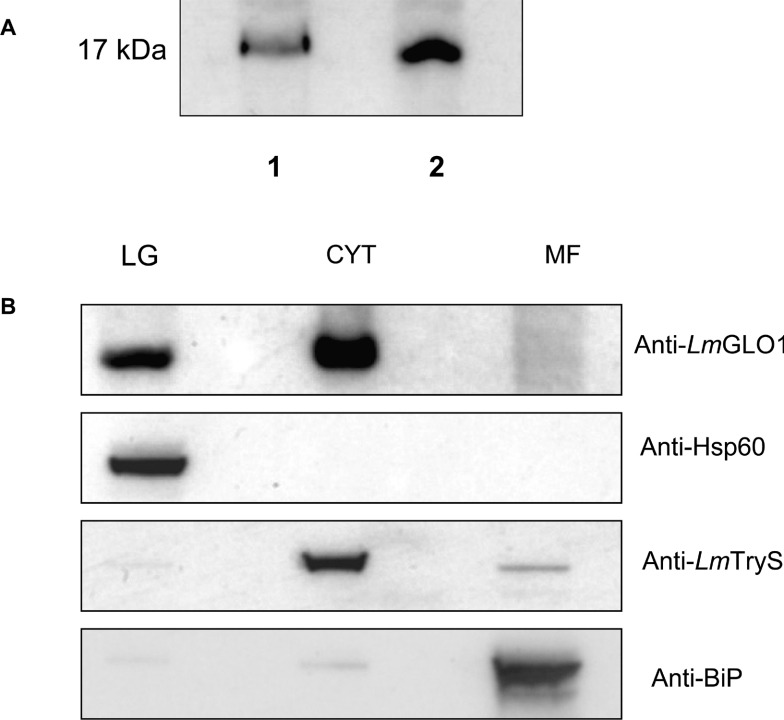
Figure 6. Immunoblot analysis of cell lysates and cellular localization of TcGLO1.
(A) Blots of whole cell extracts (30 μg of protein in each lane) from T. cruzi epimastigotes (lane 1) and L. major promastigotes (lane 2) were probed with an anti-(L. major GLO1) antiserum. (B) Subcellular fractions of L. major promastigotes, containing the large granule (LG), cytosol (C), and microsomal fractions (MF), were prepared as described in the Materials and methods section. Western blots of these equally loaded fractions (30 μg of protein in each lane) were probed with anti-(L. major GLO1) antibody. In addition, blots were stripped and reprobed with antiserum to marker proteins for each subcellular fraction to demonstrate the purity of each fraction [LG, anti-Hsp60; C, anti-(L. major trypanothione synthetase); and MF, anti-BiP].

