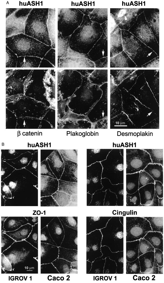
Figure 4.
huASH1 protein localizes specifically to tight junctions. (A) Caco2 cells were double-stained with guinea pig Ab against huASH1 together with mAb against the adherens junction proteins β-catenin and plakoglobin, or together with mAb against the desmosomal protein desmoplakin. Note that all junction proteins (including those in B) are localized in the vicinity of each other, being components of the subapical junctional complex. Nevertheless, the distribution of huASH1 varies from that of the other proteins. Regions with the clearest variations are indicated by arrows (see text). It is noteworthy that, in mitotic cells, huASH1 is associated with the mitotic spindle (arrow at Right Top corner). (B) IGROV1 and Caco2 cells double-stained with guinea pig Ab against huASH1 and with mAb against the tight junction proteins Z0-1 or cingulin. Here, the membranal staining patterns appear identical.

