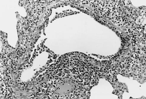
FIG. 2.
Medium-sized bronchiole from a 4-week-old pig 5 days p.i. with Sw/TX/98 virus (cluster I). The airway is lined irregularly with immature cuboidal epithelial cells. The lumen contains sloughed necrotic epithelial cells and a few neutrophils. A loose cuff of infiltrating lymphocytes surrounds the bronchiole (magnification, ×149).

