Figure 3.
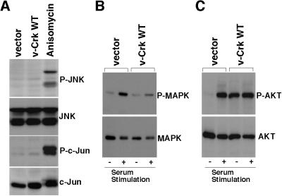
Investigation of the downstream signals of v-Crk. (A) Analysis of the JNK pathway. Total cell lysates were prepared from CEF transduced with control vector, CEF transduced with v-crk WT, and CEF treated with anisomycin (10 μg/ml for 20 min). Cell lysates then were subjected to immunoblot analysis with antibodies specific for the phosphorylated forms of either JNK (P-JNK) (first panel) or c-Jun (P-c-Jun) (third panel). The same blots also were probed with antibodies to JNK (second panel) or to c-Jun (fourth panel) that react with these proteins irrespective of phosphorylation state to confirm that equal amounts of each protein were present in each lane. (B) Analysis of the MAPK pathway. CEF transduced with vector or v-crk WT were serum-starved for 24 h and then stimulated with 20% calf serum for 20 min (+), or left unstimulated (−). Total cell lysates from these cells were subjected to immunoblot analysis with antibody specific for the phosphorylated form of MAPK (P-MAPK) (Upper). The same blots were also probed with antibody to MAPK that reacts with MAPK irrespective of phosphorylation state to confirm that equal amounts of this protein were present in each lane (Lower). (C) Analysis of the PI3K/AKT pathway. CEF transduced with vector and CEF transduced with v-crk WT were serum-starved for 24 h and then stimulated with 20% calf serum for 20 min (+), or left unstimulated (−). Total cell lysates from these cells were subjected to immunoblot analysis with antibody specific for the phosphorylated form of AKT (P-AKT) (Upper). The same blots also were probed with antibody to AKT that reacts with AKT irrespective of phosphorylation state to confirm that equal amounts of this protein were present in each lane (Lower).

