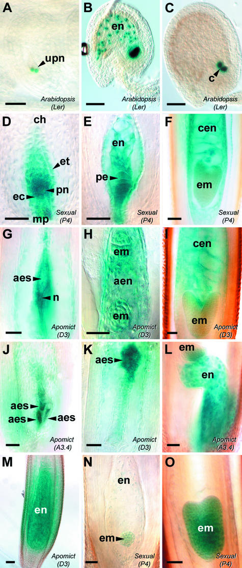
Figure 2.
AtFIS2:GUS and AtFIE:GUS Expression during Early Seed Development.
Ovules from Arabidopsis Landsberg erecta AtFIS2:GUS ([A] to [C]), sexual Hieracium P4 ([D] to [F], [N], and [O]), and apomictic Hieracium D3 and A3.4 ([G] to [M]) were stained with GUS and viewed whole-mount using Nomarski differential interference contrast (DIC) microscopy. (A) to (M) show AtFIS2:GUS expression, and (N) and (O) show AtFIE:GUS expression. Bars = 50 μm.
(A) Anthesis ovule containing unfused polar nuclei (upn) within the mature embryo sac.
(B) Postfertilization ovule containing dividing endosperm nuclei (en).
(C) Postfertilization ovule after the sixth mitotic division of endosperm development, with GUS staining in the endosperm cyst (c).
(D) Mature embryo sac in the chalazal (ch)-to-micropylar (mp) orientation surrounded by the endothelium (et), containing a fused polar nucleus (pn) and egg cell (ec).
(E) Postfertilization embryo sac containing dividing endosperm nuclei and a dividing proembryo (pe).
(F) Developing fertilized seed containing an early-heart-stage embryo (em) and cellular endosperm (cen).
(G) Mature aposporous D3 embryo sac (aes) containing central cell–like nuclei (n).
(H) Aposporous D3 embryo sac showing dividing autonomous endosperm nuclei (aen) and two globular embryos.
(I) Developing D3 seed showing a heart-stage embryo and autonomous cellular endosperm.
(J) Multiple aposporous embryo sac structures developing within a single A3.4 ovule.
(K) Displaced aposporous embryo sac developing in the chalazal region of a D3 ovule.
(L) Developing A3.4 seed containing autonomous endosperm and showing parthenogenetic embryo development at the chalazal end of the embryo sac.
(M) Late D3 seed containing autonomous cellular endosperm and no embryo.
(N) Sexual P4 embryo sac containing a globular embryo and dividing endosperm nuclei.
(O) Developing P4 seed containing an early-heart-stage embryo.

