Figure 3.
SERK-Like Genes in Hieracium.
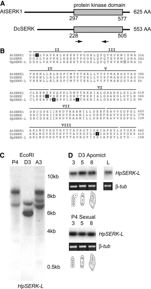
(A) Scheme of the AtSERK1 and DcSERK amino acid sequences showing the positions of the protein kinase domain and the degenerate primers (arrows). AA, amino acids.
(B) Alignment of the putative amino acid sequences for AtSERK1 and DcSERK over the length of the Hieracium SERK-like (HpSERK-L) fragment. Nonconserved residues are highlighted in black, and similar residues are highlighted in gray. The positions of the kinase domains (II to VIII) are indicated above the sequences (Schmidt et al., 1997).
(C) Genomic analysis of HpSERK-L. Genomic DNA samples were digested with EcoRI, which cuts external to the HpSERK-L fragment.
(D) RT-PCR expression analysis of HpSERK-L during ovary development. Total RNA was isolated from stages 3, 5, and 8 of ovary development, which coincide with meiosis, megagametogenesis, and early embryo development, respectively. The diagrams indicate the approximate stages of embryo sac development. RT-PCR samples were compared to a β-tubulin control (β-tub). L, leaves.

