Figure 5.
AtSPL:GUS Expression during Floral Development in Hieracium.
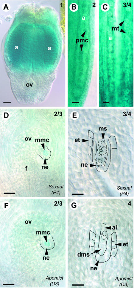
Cleared GUS-stained Hieracium florets ([A] to [C]) and ovaries ([D] to [E]) viewed whole-mount using Nomarski DIC microscopy. The numbers at top right indicate the ovary stage (Koltunow et al., 1998). Bars = 50 μm in (A) to (C) and 25 μm in (D) to (G).
(A) Early floret from apomictic D3 containing a small ovule (ov) and immature anthers (a).
(B) Enlargement of an anther from apomictic D3 containing pollen mother cells (pmc).
(C) Anthers from sexual P4 containing microspore tetrads (mt).
(D) Ovule from sexual P4 containing a megaspore mother cell (mmc) showing GUS activity surrounded by the nucellar epidermis (ne). The funiculus (f) is indicated to aid orientation.
(E) Ovule from sexual P4 showing four megaspores (ms), outlined with dashed lines, surrounded by the nucellar epidermis and the developing endothelium (et), outlined with solid lines. No staining is detected in the indicated structures.
(F) Ovule from apomictic D3, showing the corresponding stage of apomictic development to (D), containing a megaspore mother cell showing GUS activity.
(G) Ovule from apomictic D3, showing the corresponding stage of apomictic development to (E), containing an expanding aposporous initial cell (ai) chalazal to the degenerating megaspores (dms). No staining is detected in the indicated structures.

