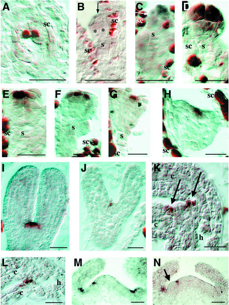
Figure 3.
In Situ Localization of CUC3 mRNA.
Sections are frontal and longitudinal unless stated otherwise.
(A) Mature embryo sac, transverse section through the polar nuclei of the central cell.
(B) Two-celled embryo (asterisks indicate the two embryo cells, and the arrow indicates the cell wall separating them).
(C) Octant-stage embryo (the arrow indicates the O line separating the upper and lower tiers).
(D) Early globular embryo.
(E) Globular embryo.
(F) Globular embryo (sagittal section).
(G) Triangular-stage embryo.
(H) Heart-stage embryo.
(I) Torpedo-stage embryo (central section through the SAM).
(J) Torpedo-stage embryo (superficial section through the boundary of the cotyledons).
(K) Mature embryo (central section through the SAM; arrows indicate the boundaries between the SAM and the cotyledons).
(L) Mature embryo (section through the boundary of the cotyledons).
(M) pt-1 seedling apex at 5 days after germination.
(N) pt-1 seedling apex at 5 days after germination with emerging leaf primordium (arrow).
c, cotyledon; h, hypocotyl; s, suspensor; sc, seed coat. Bars = 20 μm.

