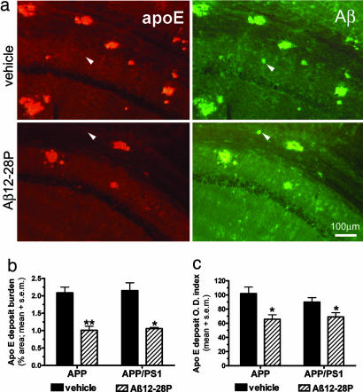
Fig. 4.
Treatment with Aβ12-28P reduces the amount of apoE present in Aβ deposits. (a) Shown is double immunofluorescent staining colocalizating apoE in Aβ deposits in the hippocampus of APPK670N/M671L mice. Only a minority of plaques were apoE-negative in both groups (see white arrowheads). (b) Shown is a reduction in the burden of apoE-positive deposits in Aβ12-28P-treated animals (∗, P < 0.01; ∗∗, P < 0.001). Values are averaged for all three areas of interest. (c) Shown is the reduction in the mean optic density (O.D.) index of apoE deposits in Aβ12-28P-treated animals (∗, P < 0.05).

