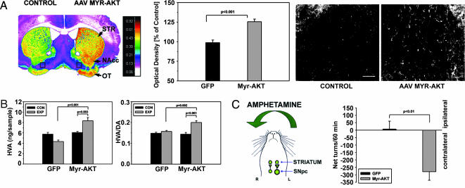
Fig. 2.
Trophic effects of Myr-Akt on the axonal projections of SN DA neurons. (A) (Left) A pseudocolor image of a coronal section through the striatum, processed for TH immunostaining at 7 weeks after intranigral AAV Myr-Akt injection, reveals higher density values (red-orange) in the striatum (STR) on the injected side. Higher-density values also are observed in the nucleus accumbens (NAcc) and olfactory tubercle (OT). (Center) An increase in the optical density of the striatal TH immunostaining is shown quantitatively as a 26% increase over values for the contralateral, noninjected striatum; this was a highly significant increase in comparison to AAV GFP-injected animals (P < 0.001, t test; n = 6 animals for both groups). (Right) At a cellular level, this increase in optical density was attributable to an increase in the number and caliber of TH-positive fibers. (Bar: 50 μm.) The regions shown in these micrographs are between the accumbens and olfactory tubercle on both sides of the section (indicated as black rectangles in Left). (B) These morphologic changes were accompanied by biochemical changes indicative of increased DA release in the striatum. There was an increase in both HVA (P < 0.001) and the HVA/DA ratio (P = 0.002, ANOVA, AAV Myr-Akt-injected side compared with AAV GFP-injected side; n = 6 animals in each group) (EXP, experimental, injected side; CON, control, noninjected side). (C) (Left) When AAV Myr-Akt-injected mice were administered amphetamine (5 mg/kg i.p.), they exhibited rotational behavior contraversive to the side of the vector injection. (Right) Contraversive rotations are plotted as negative net rotations. AAV GFP-injected mice did not demonstrate such behavior.

