Figure 6.
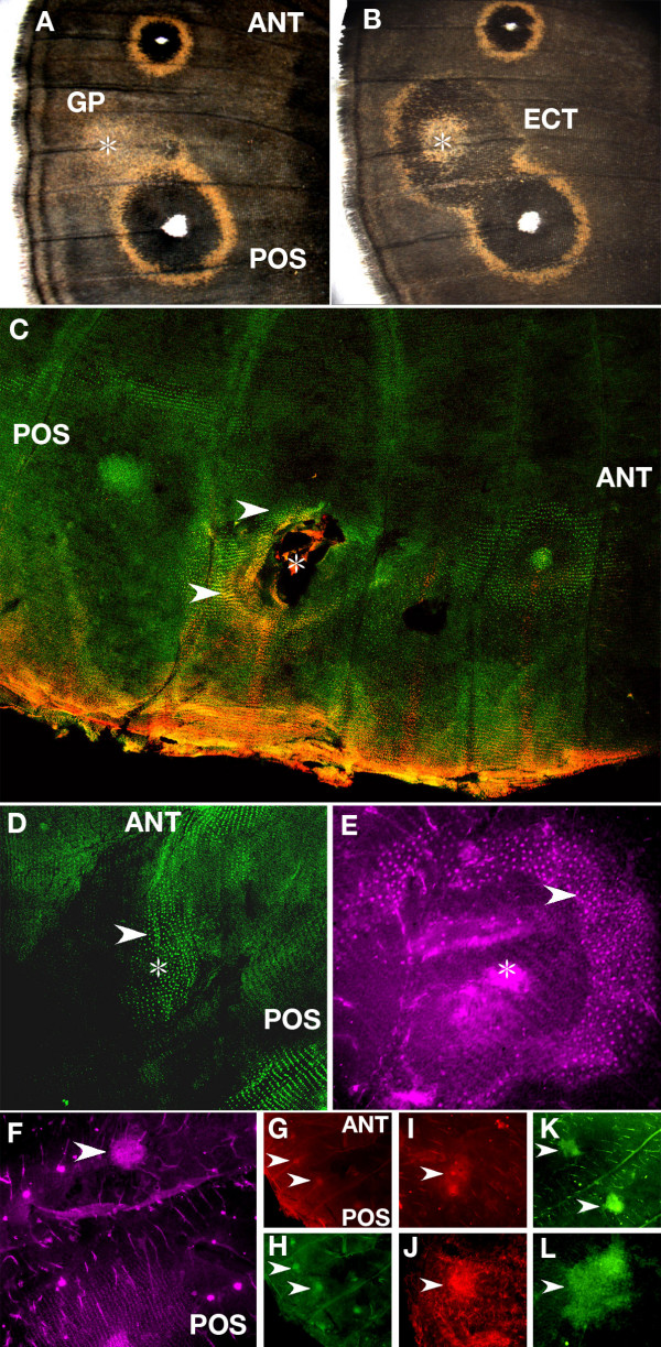
Gene expression after epidermal wounding in Bicyclus anynana. (A) Gold patches (GP) of scales are produced in some individuals around a wound site (star symbol) between the anterior (ANT) and posterior eyespots (POS), whereas (B) ectopic eyespots (ECT) containing both black scales and gold scales differentiate in other individuals; (C) Dll (red) and En (green) are present in scale-building cells around a wound site (yellow represents co-expression). Epidermis was wounded at 12 h after pupation (W = 12 h) followed by tissue fixation and protein visualization at 25 h after pupation (V = 25 h) (50×); (D) en is expressed in a patch of scale-building cells (arrowhead) around wound site (star symbol) (W = 9 h, V = 24 h) (100×); (E) sal is expressed in scale-building cells (arrowhead) around a wound site (approximate location shown by star symbol) (W = 13.5 h, V = 28.5 h) (200×). The absence of a continuous epidermis in the center of the wound site in (C) and (E) is the result of damage during the process of wing detachment from the overlying cuticle due to the presence of a wound scab; (F-L) Antibodies bind to the center of a wound (shown in all panels by arrowheads) in a non-specific fashion; (F) anti-Sal antibody binds to a cluster of non-differentiated cells (W = 11.5 h, V = 24 h) (100×); (G) Anti-Dll (W = 9 h, V = 24 h) (50×); (H) Anti-En (W = 9 h, V = 24 h) (50×). Note the two flanking eyespot foci on this wing (G, H) expressing Dll and en; (I) Anti-Wg (W = 9 h, V = 24 h) (100×); (J) Anti-pSmad (W = 9 h, V = 24 h) (100×); (K) Control staining using only an anti-mouse secondary antibody (and no primary antibody) also showing expression at the center of two wound sites (W = 9 h, V = 24 h) at 50X, and at 200X (L).

