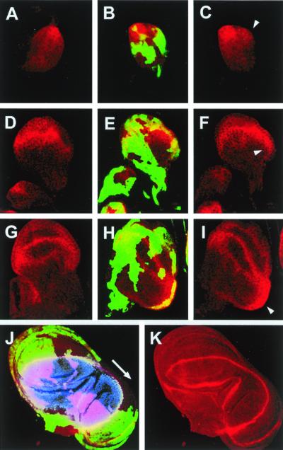
Figure 3.
DER signaling antagonizes Wg expression. DNRaf3.1-expressing cells were identified by the expression of green fluorescent protein (green). Wg expression is shown in red. (A, D, and G) Control wild-type discs. (B, C, E, F, H, and I) Discs with clones of cells that overexpressed DNRaf3.1 under the control of actin-Gal4 (see Materials and Methods). (A–C) Early L2 wing discs. Wg is expressed in the anterior-ventral quadrant. In the absence of DER signaling, Wg expression is induced in a more posterior position (arrowhead). (D–F) Late L2 wing discs. Wg is expressed in the presumptive wing pouch. Wg expression extends to posterior proximal positions (arrowhead) in discs lacking DER signaling. (G–I) Early L3 wing discs. Expression of Wg evolves in the wing margin and wing pouch circles. In the absence of DER signaling, Wg expression expands further to posterior proximal positions (arrowhead). (J and K) Late L3 wing disk. Wg is ectopically expressed in the duplicated wing pouch. Nuclear apterous expression (blue) outlines the expansion of the wing dorsal compartment to the duplicated wing pouch, stretching out the D/V border to more posterior cells.

