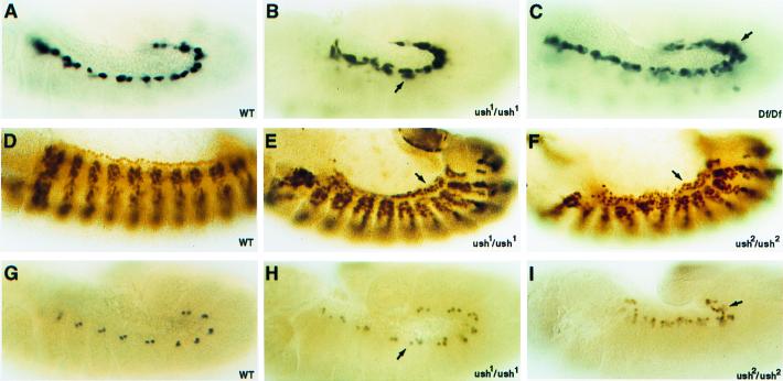
Figure 2.
ush gene functions during Drosophila cardiogenesis. (A, D, and G) WT, wild-type embryos. (B, C, E, F, H, and I) Homozygous ush embryos of the indicated genotype. (A–C) Lateral views of stage 12 embryos stained for lacZ reporter gene expression under the control of a D-mef2 cardial cell enhancer. (D–F) Lateral views of stage 13 embryos stained for D-MEF2 protein in cardial cells. (G–I) Lateral views of stage 12 embryos stained for Eve protein in a subset of pericardial cells. Arrows point to the increased numbers of cardial or pericardial cells in the ush mutant embryos.

