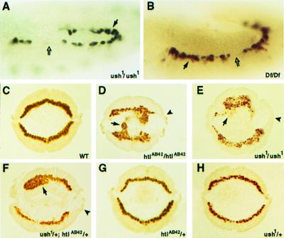
Figure 5.
ush function in mesodermal cell migration. (A and B) Lateral views of stage 11 and 12 embryos of the genotypes ush1/ush1;IIA341–44/+ and Df(2L)al/Df(2L)al;IIA341–44/+, respectively, stained for β-galactosidase produced in cardial cells under the control of a D-mef2 heart enhancer. Solid arrows point to increased numbers of cardial cells whereas open arrows indicate the absence of cells in some hemisegments. (C–H) Analysis of mesodermal cell migration defects. Stage 10 embryos of the indicated genotypes were stained for D-MEF2 protein and sectioned to visualize the normal or abnormal spreading of the mesodermal cell layer. Arrows point to clusters of mesodermal cells, whereas arrowheads indicate the dorsal-most ectodermal cells that correspond to the normal position of cell spreading in wild-type embryos. Strong migration defects are observed in homozygous htl and ush mutants and in embryos transheterozygous for the two genes.

