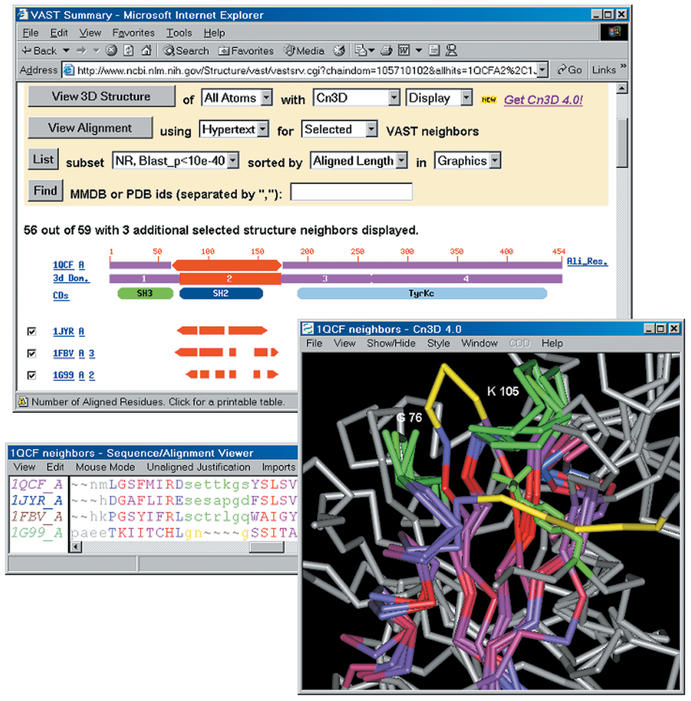
Figure 2.
MMDB's ‘VAST summary’ of selected structure neighbours of the SH2 domain (3D domain 2) of Hck kinase/1QCF. The locations of loop regions whose conformation is conserved in Grb2/1JYR and c-Cbl/1FBV are highlighted in green, as is the phosphotyrosine residue of a peptide bound to 1FBV. Analogous loop regions in acetate-kinase/1G99 are highlighted in yellow. The loop analogous to that near 1QCF K105 adopts a different conformation and the loop analogous to that near 1QCF G75 occludes the site where SH2 domains bind phosphotyrosine. Cn3D's alignment window displays the residues of 1QCF that may be superposed onto all selected neighbours; the degree of sequence conservation is indicated via a colour-ramp from blue-grey to red, with non-aligned sites shown in grey.

