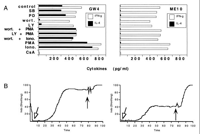
Figure 1.
Discordant expression of PI3-kinase-regulated events differentiates IL-4+ from IL-4-null Vα24JαQ T cell clones. (A) Vα24JαQ T cell clones GW4 (nondiabetic) and ME10 (diabetic) were activated with plate-bound anti-CD3 or Ig control. Levels of secreted IL-4 and IFN-γ were assayed by ELISA. The concentrations of inhibitors used were 10 nM wortmannin (wort.); 10 μM LY294002 (LY); 50 μM PD98059 (PD), a mitogen-activated protein kinase kinase inhibitor; and 50 μM SB203580 (SB), a p38 kinase inhibitor. Data points were collected in triplicate, and the Fig. is representative of four independent experiments. The concentrations of phorbol ester and calcium ionophore used were 1 ng/ml PMA and 1 μg/ml ionomycin (Iono). Cyclosporin A (CsA) was used at 5 ng/ml. (B) Vα24JαQ T cell clones GW4 (IL-4+) and ME10 (IL-4-null), 1 × 10⁶ cells each, were loaded with Indo-1 (10 μM) for 45 min, then stimulated with 10 μg/ml anti-CD3 (open arrowhead) and analyzed on a Cytomation MoFlo instrument. At the end of the experiment, ionomycin was added to a final concentration of 1 μg/ml (filled arrowhead) to determine maximal flux. The ratio of Indo-1 fluorescence 410/490 nm (410, Ca2+-bound; 490, Ca2+-free) after stimulation in a representative pair of clones is pictured. Control experiments showed no differences in calcium flux in response to thapsigargin treatment (data not shown).

