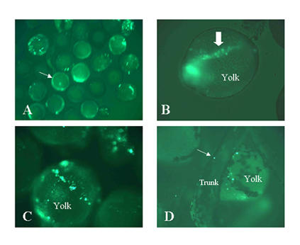
Figure 8.

Expression of promoter 1a fusion construct: labelling of yolk platelets. The exon 1a constructs shown in the upper panel of figure 6 were injected into zebrafish embryos at the 1–4 blastomere stages. Live embryos were observed at various stages of development under a fluorescent microscope to detect the EGFP-F reporter. Panel A: low magnification view of multiple embryos 10 hrs after fertilization (1–5 somite stages). The speckles are yolk platelets labeled by EGFP-F. They are slightly out of focus due to bulk motion of the water in which the embryos are suspended. The arrow points to a developing embryo on top of the yolk. The embryo exhibits background fluorescence. Panel B: A 10 hrs old embryo in which some of the yolk platelets line up with the developing trunk and head. The arrow points to the trunk. The head, to the left, is out of focus. Panel C: 10 hrs old embryo. The line is not coincident with the body axis in this embryo. Panel C: 2 days old embryo. Some platelets remain in the yolk. The arrow points to a platelet in the trunk.
