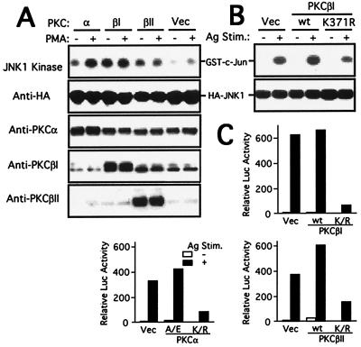
Figure 3.
PKCβI regulates the JNK pathway and cytokine gene expression. (A) COS-7 cells were transfected with HA-JNK1 plasmid together with an empty vector (vec), wt PKCα, wt PKCβI, or wt PKCβII vectors. Forty-eight hours later, cells were left unstimulated or stimulated with 100 nM PMA for 10 min. Cell lysates were immunoprecipitated with anti-HA, and immunoprecipitates were incubated with GST-c-Jun (1–79) in the presence of [γ-32P]ATP. Reactions were analyzed by SDS/PAGE and autoradiography (Top). Expression of HA-JNK1, PKCα, PKCβI, or PKCβII was detected by immunoblotting of total cell lysates. (B) Bone marrow-derived mast cells from wt mice were transfected with wt or kinase-dead (K371R) PKCβI cDNAs together with HA-JNK1 plasmid. Forty-eight hours later, cells were stimulated with IgE and antigen. Anti-HA immunoprecipitates were subjected to kinase assays. (C) Wt mast cells were transfected with IL-2Luc plasmid (8 μg) together with wt or mutant cPKC cDNAs (20 μg each). Cells were stimulated with IgE and antigen for 8 h. Luciferase activity was measured. PKCα A/E is a constitutively active form of PKCα. Similar data were obtained with TNF-αLuc plasmid (not shown).

