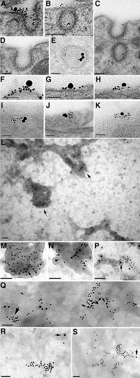
Fig. 3. PrPC immunolabelling of sensory neurons, seen by transmission electron microscopy of sectioned material (A–K) or of tear-off samples of the cell surface (L–S), using anti-PrP Fab directly coupled to 5 nm gold with biotinylated Tf coupled to 20 nm avidin–gold (A–K) or AP2 labelled with 10 nm gold (L–S). (A–E) Examples of PrPC entering coated pits or vesicles after 2 min at 37°C, with occasional co-localization with TfR (A and E). (F–K) Co-localized TfR and PrP on the cell surface. (L) Overlapping PrP and AP2 at electron-dense cytoplasmic tubular structures (M and N) Regions arrowed in (L) at higher power; arrows in (M) and (N) point to 10 nm AP2 label. (P) A cluster of PrPC partially overlapping an AP2-labelled electron-dense region (arrowed). (Q) Clusters of PrPC overlying AP2 (small arrows), with another PrPC cluster (large arrow) not associated with AP2. In this sample, the cytoplasmic surface is less distinct (presumably masked by cytoplasmic protein), somewhat obscuring the AP2-positive electron-dense structures. (R and S) Examples of clusters of PrP not associated with AP2 label (the nearest AP2 label is arrowed). Scale bars are 50 nm.

An official website of the United States government
Here's how you know
Official websites use .gov
A
.gov website belongs to an official
government organization in the United States.
Secure .gov websites use HTTPS
A lock (
) or https:// means you've safely
connected to the .gov website. Share sensitive
information only on official, secure websites.
