
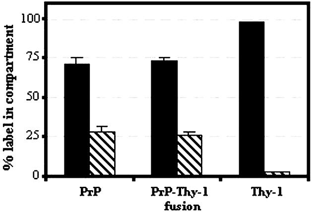
Fig. 8. Distribution of 5 nm gold–Fab detecting PrP, PrP107–Thy-1 (Fusion) and Thy-1 on the cell surface (black) and within intracellular vesicles (stripes) on N2a cells after 10 min incubation at 37°C. The percentages of labelled PrPC and fusion protein on the cell surface and within intracellular vesicles were similar (P = 0.57 and 0.64, respectively) and differed from Thy-1 (P < 0.0001).