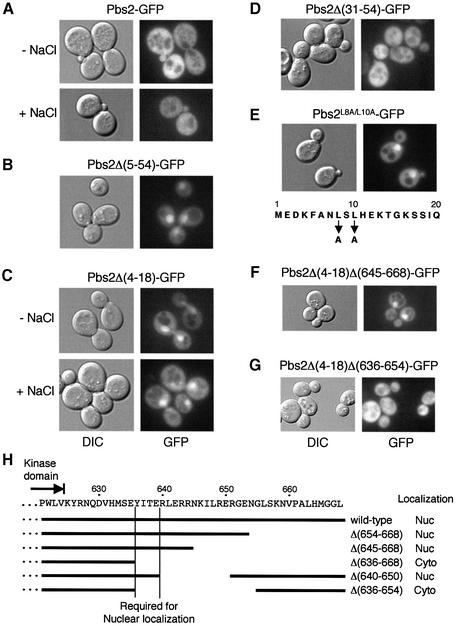
Fig. 6. Subcellular localization of the wild-type and mutant Pbs2 proteins. (A–G) TM261 (pbs2Δ) was transformed with either a plasmid expressing the wild-type Pbs2 fused to GFP (Pbs2–GFP) or its mutant derivatives, as indicated. Cells were grown to mid-log phase in CAD medium, and localization of the GFP fusion proteins was determined by fluorescence microscopy as described in Materials and methods. Localization of Pbs2–GFP and Pbs2Δ(4–18) was examined before (–NaCl) and 5 min after (+NaCl) addition of 0.4 M NaCl. Localization of the other proteins was observed in the absence of osmotic stress. The nuclear export signal (NES), within the first 20 amino acid residues of Pbs2, is shown in (E). DIC, differential interference contrast. (H) Analysis of Pbs2 C-terminal region deletion mutants. The Pbs2 C-terminal amino acid sequence, with the end of the kinase domain indicated by an arrow, is shown above. Below the sequence, C-terminal deletion mutations are indicated by horizontal bars. Thease deletion mutations were combined, individually, with the N-terminal NES mutation Δ(4–18), and the subcellular localizations of the double mutants were examined as in (F) and (G). ‘Wild-type’ refers to the Δ(4–18) mutation alone. Nuc, nuclear localization; Cyto, cytoplasmic localization.

An official website of the United States government
Here's how you know
Official websites use .gov
A
.gov website belongs to an official
government organization in the United States.
Secure .gov websites use HTTPS
A lock (
) or https:// means you've safely
connected to the .gov website. Share sensitive
information only on official, secure websites.
