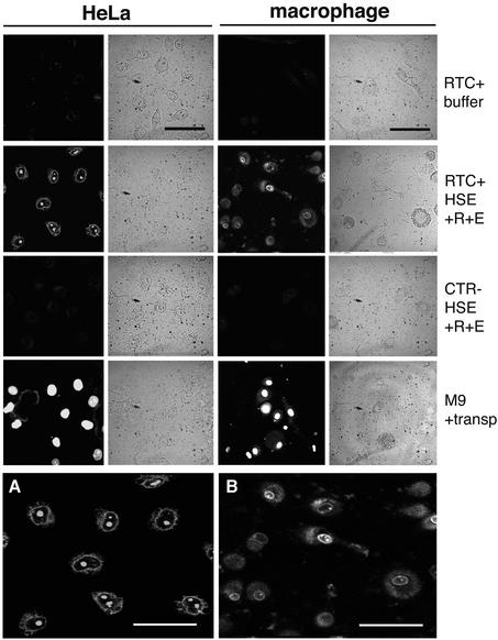
Fig. 2. Nuclear import assay with labelled RTCs in HeLa cells and primary human macrophages. Following permeabilization with digitonin, cells were incubated with 103 RTCs/cell (RTC+) or an equal volume of sample from uninfected cells (CTR–) or 1 µM fluorescent-labelled M9-bearing nucleoplasmin core (M9) plus transportin (transp, 1 µM). Incubation was performed for 15 min at 37°C in the presence of energy mix (E), Ran mix (R) and HeLa high-speed cytosolic exctracts (HSE, 0.5 mg/ml final concentration) or buffer. Fluorescent and transmission images of the same fields are shown. Scale bars, 25 µm. (A) Enlarged image corresponding to the HeLa RTC HSE panel. (B) Enlarged image corresponding to the macrophage RTC HSE panel. Scale bars, 25 µm.

An official website of the United States government
Here's how you know
Official websites use .gov
A
.gov website belongs to an official
government organization in the United States.
Secure .gov websites use HTTPS
A lock (
) or https:// means you've safely
connected to the .gov website. Share sensitive
information only on official, secure websites.
