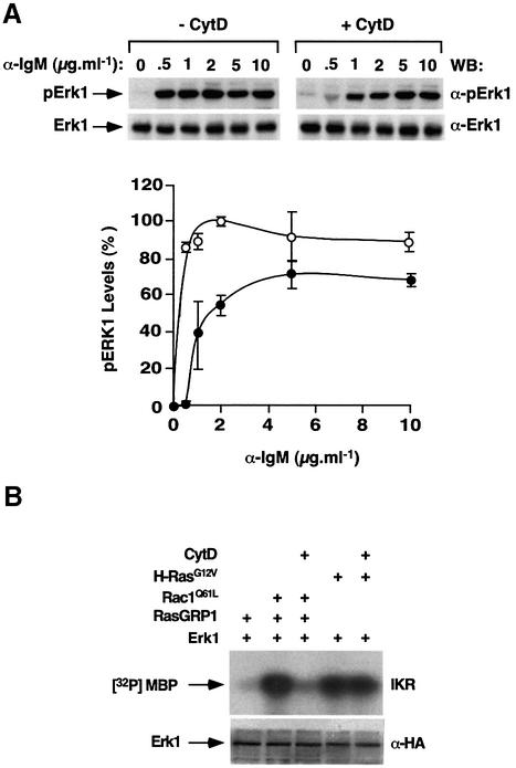
Fig. 7. (A) Effect of cytochalasin D on the activation of the Ras cascade in DT40 cells. Exponentially growing cells were cultured for 2 h in the absence (–CytD) or presence (+CytD) of cytochalasin D (10 µM) and then stimulated for 10 min with the indicated amounts of anti-IgM antibodies. Cells were then lysed and the resulting extracts analyzed by immunoblot analysis using either anti-phosphoErk1 (top panels) or anti-Erk1 (bottom panels) antibodies. The graph shows the mean and SD values of Erk1 activation obtained in three independent experiments (100% is the maximal value of activation of Erk1). (B) Effect of cytochalasin D on the Vav/Rac–RasGRP1 interaction in COS1 cells. COS1 cells expressing HA-Erk1 in the presence of the indicated proteins were incubated with cytochalasin D for 1 h before evaluating the kinase activity of Erk1 using in vitro kinase reactions (upper panel). Cell lysates from the same experiments were analyzed in parallel by anti-HA immunoblots to evaluate the expression levels of Erk1 under each experimental condition (lower panel).

An official website of the United States government
Here's how you know
Official websites use .gov
A
.gov website belongs to an official
government organization in the United States.
Secure .gov websites use HTTPS
A lock (
) or https:// means you've safely
connected to the .gov website. Share sensitive
information only on official, secure websites.
