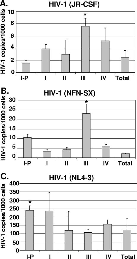
FIG. 5.
HIV-1 entry in thymocyte subsets at different stages of maturation. Postnatal thymocytes were separated into individual subsets by magnetic beads and infected in vitro. Eighteen hours postinfection, quantitative PCR was performed to measure the number of initial reverse transcription products (R-U5), and the cell numbers were determined by beta-globin quantitative PCR. Values were calculated based on interpolation from a standard curve, and HIV-1 copy numbers were normalized to 1,000-cell equivalents from each subset after infection by R5 HIV-1JR-CSF (A), R5 HIV-1NFN-SX (B), and X4 HIV-1NL4-3 (C). The mean viral-copy numbers and standard deviations for X4 and R5 HIV-1 DNA based on triplicate measurements in one representative experiment out of three are shown. Asterisks indicate statistically significant differences calculated based on data from three independent experiments.

