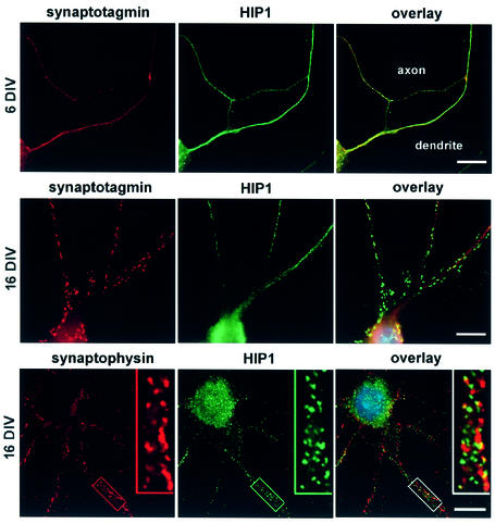
Fig. 6. HIP1 is expressed in the somatodendritic compartment in primary hippocampal neurons. HIP1 expression was analyzed by double immunofluorescence in primary hippocampal neurons after 6–21 days in culture. Synaptotagmin and synaptophysin immunolabeling is shown in red. HIP1 immunostaining was either detected with the mAb HIP1#9 (top and middle) or the pAb HIP1FP (bottom) and is shown in green. HIP1 does not colocalize with synaptic vesicle markers and shows a greater enrichment in the somatodendritic compartment. Nuclei were counterstained with DAPI shown in the electronic overlays (in blue). Scale bar = 10 µM.

An official website of the United States government
Here's how you know
Official websites use .gov
A
.gov website belongs to an official
government organization in the United States.
Secure .gov websites use HTTPS
A lock (
) or https:// means you've safely
connected to the .gov website. Share sensitive
information only on official, secure websites.
