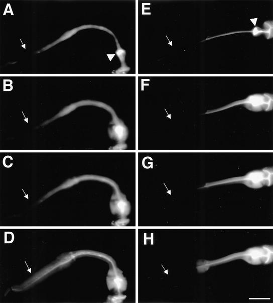
Figure 6.
Cell-cell coupling is abnormal in the pharyngeal muscle of inx-6(rr5) mutants. Wild-type N2 (A) and inx-6(rr5) mutant animals (E) were allowed to ingest a saturated carboxyfluorescein solution. Dye was accumulated in the pharyngeal lumen and was selectively excluded from the pharyngeal muscle in wild-type and inx-6(rr5) animals but could enter the muscle and diffuse anteriorly after a single laser pulse focused at the posterior of the grinder. (A–D) Dye-coupling process in the wild-type L1 stage animal, showing diffusion of the fluorescent dye toward the procorpus after the laser pulse. (E–H) Same process in the inx-6(rr5) L1 mutant preincubated at restrictive temperature. Arrowheads indicate the grinder in the terminal bulb where the laser pulse was applied. White arrows indicate the procorpus. In wild-type animals, dye spreads anteriorly through the isthmus and throughout the whole corpus evenly within 60 s after laser treatment, whereas in inx-6(rr5) animals, dye quickly spreads into the metacorpus but could not cross into the procorpus. Images were captured within 60 s after laser treatment by using the same exposure time for each acquisition. The final dye-coupling pattern reached equilibrium and did not change as verified 10 min after the initial laser application. Bar, 10 μm.

