Figure 7.
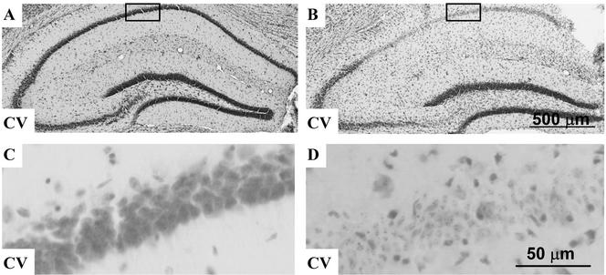
Infusion of anti-laminin γ1 antibody promotes neurodegeneration in tPA-deficient mice. TPA-deficient mice were infused with anti-laminin γ1 antibody (0.2 mg/ml) for 5 d (A and B) and then kainate was injected (B). Two days after kainate injection, the mice were killed and brain sections were prepared. The brain sections were processed for cresyl violet staining (A and B). Infusion of anti-laminin γ1 antibody was not neurotoxic by itself (A and C), but when combined with kainate injection promoted neurodegeneration in tPA-deficient mice (B and D). Higher magnification of the boxed areas in A and B are shown in C and D, respectively. CV, cresyl violet staining.

