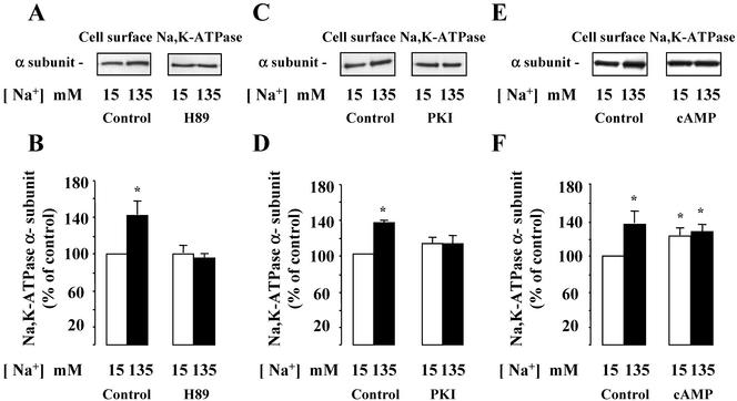
Figure 6.
Effect of PKA inhibition and activation on [Na+]i-dependent Na,K-ATPase cell surface expression of in mpkCCDcl4 cells and Na,K-ATPase activity in isolated rat CCDs. (A and B) Confluent mpkCCDcl4 cells grown on polycarbonate filters were first preincubated in the presence of either 15 or 135 mM Na+ without or with 5 × 10⁻⁵ M H-89 for 30 min at 37°C. After permeabilization by 1 μg/ml amphotericin B, cells were incubated for 1 h at 37°C. Cell surface Na,K-ATPase was detected as described in the legend of Figure 2. (A) Representative immunoblot showing Na,K-ATPase cell surface expression. (B) Bars represent densitometric values expressed as the percentage of the optical density value measured in the presence of 15 mM Na+. Results are means ± SE from four independent experiments. *p <0.05 vs. 15 mM Na+ values. (C and D) Confluent mpkCCDcl4 cells grown on polycarbonate filters were first preincubated in the presence of either 15 or 135 mM Na+ without or with 50 μM myristoylated PKI for 30 min at 37°C. After permeabilization by 1 μg/ml amphotericin B, cells were incubated for 1 h at 37°C. Cell surface Na,K-ATPase was detected as described in the legend of Figure 2. (A) Representative immunoblot showing Na,K-ATPase cell surface expression. (B) Bars represent densitometric values expressed as the percentage of the optical density value measured in the presence of 15 mM Na+. Results are means ± SE from four independent experiments. *p < 0.05 vs. 15 mM Na+ values. (E and F) Confluent mpkCCDcl4 cells grown on polycarbonate filters were first preincubated in the presence of 15 or 135 mM Na+ for 30 min at 37°C. After permeabilization by 1 μg/ml amphotericin B, cells were incubated for 1 h at 37°C and 10–3 M db-cAMP was added or not for the last 10 min of incubation. Cell surface Na,K-ATPase was detected as described in the legend of Figure 2. (D) Representative immunoblot showing Na,K-ATPase cell surface expression. (E) Bars represent densitometric values expressed as the percentage of the optical density value measured in the presence of 15 mM Na+. Results are means ± SE from four independent experiments. *p < 0.05 vs. 15 mM Na+ values.

