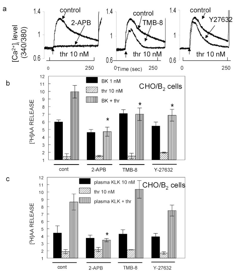
Fig 3. Role of Gα12/13 in potentiation.

Panel a: Inhibition of thrombin-induced [Ca2+]i release. Fura-2AM loaded CHO/B2 cells were incubated with 75 μM 2-APB, 20 μM TMB-8 or 10 μM Y27632 for 30 min to inhibit IP3 R, [Ca2+]i release or RhoA kinase. Cells were then exposed to 10 nM thrombin. Abscissa: time in seconds. Ordinate: relative [Ca2+]i level. Experiments were repeated three times with similar results. Panels b and c: [3H]AA loaded CHO/B2 cells were treated with 75 μM 2-APB, 20 μM TMB-8 or 10 μM Y27632 for 30 min at 37 ºC and then exposed to 1 nM BK (panel b) or 10 nM plasma kallikrein (panel c) with or without 10 nM thrombin. Solid column = agonist alone; diagonal lines = PAR1 agonist alone; vertical lines = combination B2R agonist and PAR1 agonist. Data are means ± S.E.M. from 3–5 separate experiments. * P < 0.03 comparing B2R agonists in thrombin stimulated control cells. 2-APB blocks Ca2+ entry into cells, the two other agents reduced it. Inhibitors abolished or reduced potentiation by thrombin. TMB-8 blocked potentiation of BK but not that of kallikrein.
