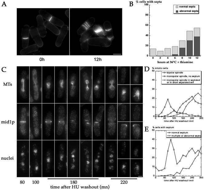
Figure 10.
Septation defects in pnmt*-E147Kcdc31 cells. (A and B) Accumulation of abnormal septa under restrictive conditions in pnmt*-E147Kcdc31 strain (AP388). Septation was monitored 2–12 h after shift to 36°C in presence of thiamine. Misorganized and multiple septa accumulate from 8 h on. Bar in A, 5 μm. (C–E) Synchronized pnmt*-E147Kcdc31 cells proceed to septation before mitosis exit. pnmt*-E47Kcdc31 mutant (AP388) was synchronized by growth for 3 h at 36°C in presence of thiamine and HU. On HU washout, cells were further incubated at 36°C in presence of thiamine, fixed, and stained for MTs, mid1p, and DNA at 20-min intervals (C and D) or stained for septum at same time points (E). In D, the percentage of cells with a bipolar spindle (squares), of nonseptated cells with a monopolar spindle (circles), or of septated cells or short cells after sister cell separation containing a monopolar spindle (diamonds) was determined according to MTs and mid1p staining. The percentage of cells presenting normal (squares) or abnormal and multiple septa (diamonds) is represented in E. Cells undergo a first normal round of mitosis (t = 80 min) and cytokinesis (t = 100 min). In the second round of mitosis, cells assemble monopolar spindles and proceed to septation 20–40 min later, before spindle breakdown (t = 180 and t = 220). Note the strong mid1p staining in the septum region in septated cells or at the new end in short separated cells containing monopolar mitotic spindles (right cells at t = 180 and bottom cells at t = 220). Note also the two supernumerary mid1p rings in the left cell at t = 220 min on each side of the septum. Bar in C, 2 μm.

