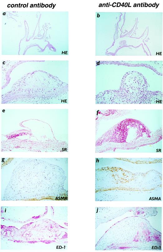
Figure 2.
Histological characteristics of delayed anti-CD40L antibody treatment. (a and b) Hematoxylin-and-eosin-stained longitudinal section of the aortic arch, including the brachiocephalic trunk, left carotid, and left subclavian artery (×25). Neither lesion area nor the number of lesions differed between anti-CD40L antibody- and control-treated mice. (c and d) Advanced atherosclerotic lesion, containing a lipid core and a fibrous cap. The relative lipid core area is less, and relative fibrous cap thickness is increased in anti-CD40L antibody- (d) compared to control-treated mice (c). (e and f) Sirius red staining of advanced atherosclerotic lesions, showing a higher relative collagen content in the anti-CD40L antibody-treated mouse (f) than in the control-treated mouse (e). (g and h) ASMA staining of advanced atherosclerotic lesions, showing a higher relative VSMC/myofibroblast content in the anti-CD40L-treated mouse (h) than in the control-treated mouse (g). (i and j) ED-1 staining of advanced atherosclerotic lesions, showing a decreased relative macrophage content in the anti-CD40L-treated mouse (j) compared to the control-treated mouse (i).

