Figure 2.
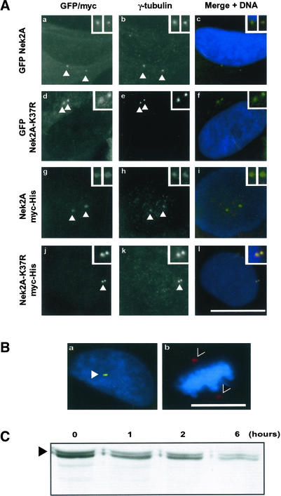
Recombinant Nek2A localizes to centrosomes but not spindle poles. Cell lines were induced with doxycycline for 24 h before processing for fluorescence microscopy. GFP-Nek2A (a–c), GFP-Nek2A-K37R (d–f), Nek2A-myc-His (g–i), and Nek2A-K37R-myc-His (j–l). GFP signal (a and d), α-myc antibodies (g and j), anti-γ-tubulin antibodies (b, e, h, and k), and merged image of GFP or myc (green), γ-tubulin (red), and Hoechst 33258 (blue) (c, f, i, and l). Bar, 15 μm. Centrosomes (arrowheads) are shown at increased magnification in insets. (B) Merged images showing Nek2A-K37R-myc-His–induced cells in interphase (a) or metaphase (b) stained with antibodies against γ-tubulin (red) and myc (green). DNA is stained in blue. Recombinant Nek2A is detected at interphase centrosomes (arrowhead) but not mitotic spindle poles (>). Bar, 15 μm. (C) Western blot with anti-Nek2 antibodies of extracts from cells induced to express Nek2A-K37R-myc-His (arrowhead) for 24 h before addition of cycloheximide for the times indicated (hours).

