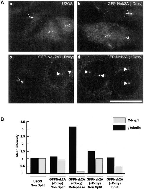
Figure 4.
C-Nap1 is still present on Nek2A-induced split centrosomes. (A) U2OS T-REx cells (a) or GFP-Nek2A cells without (b) or with 24-h doxycycline induction (c and d) were fixed and immunostained with anti-C-Nap1 antibodies. Images were captured under identical conditions to allow comparison of C-Nap1 signal intensity. Unsplit (>) and split (filled arrowheads) centrosomes in interphase centrosomes, and mitotic spindle poles (open arrowheads) are indicated. Bar, 15 μm. (B) Abundance of C-Nap1 and γ-tubulin at centrosomes was calculated by measuring mean pixel intensities as indicated in MATERIALS AND METHODS. Centrosomes in 20 cells for each condition were imaged, quantitated, and normalized to the intensity of unsplit interphase centrosomes in the parental U2OS cell line (given an arbitrary value of 1.0). Results show the mean of three independent experiments in which all cells were fixed at the same time and immunostained with C-Nap1 antibodies under identical conditions.

