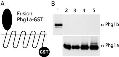
Figure 7.
No detectable interaction between Phg1a and Phg1b proteins. (A) PHG1a knockout cells were transfected with a construct encoding Phg1a C-terminal fusion to GST tagged with a FLAG epitope, Phg1a-GST. (B) Triton X-100 (lanes 2 and 3) or CHAPS (lanes 4 and 5) lysates from cells expressing the Phg1a-GST fusion were incubated with Sepharose beads coupled to glutathione (lanes 2 and 4) or to an antibody against the FLAG epitope (lanes 3 and 5). The adsorbed proteins were processed for Western blotting and revealed with an antiserum to the Phg1b protein (top) or the Phg1a protein (bottom). To indicate the position of the Phg1b protein, an aliquot of lysate from a cell overexpressing Phg1b was comigrated (lane 1).

