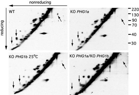
Figure 8.
Pattern of cell surface proteins is altered in PHG1 knockout cells. After cell surface biotinylation, cells were lysed and total proteins resolved on a two-dimensional nonreduced/reduced gel. Biotinylated proteins were revealed after incubation with horseradish peroxidase-avidin conjugate. The cell surface expression of at least three proteins (indicated by arrows) was markedly reduced in mutants exhibiting a phagocytosis defect.

