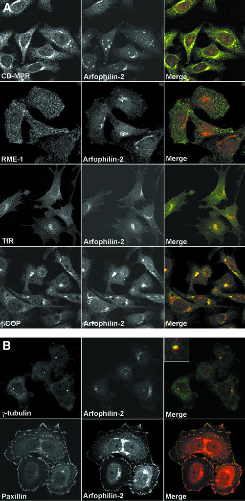
Figure 3.
Intracellular localization of endogenous arfophilin-2. (A) Antibodies specific for arfophilin-2 were used to localize the endogenous protein in HeLa cells (middle, red in merged images) and to compare its distribution with marker proteins (left, green in merged images). Arfophilin-2 staining was predominantly detected in a perinuclear region partially coincident with the CI-MPR and βCOP but with little overlap with the TfR or RME-1. (B) Minor arfophilin-2 staining (red in the merged images) in the centrosome region, adjacent to γ-tubulin, and also in focal adhesions where it colocalized with paxillin. Data are typical of eight experiments. Note that to reveal the staining at focal adhesions, significantly increased laser power was used to collect this image with respect to that shown in A.

