
Figure 2.
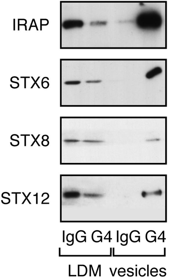
Colocalization of t-SNAREs in Glut4 vesicles. GLUT4 vesicles were isolated from 3T3-L1 adipocyte postnuclear supernatants as described in text. anti GLUT4 (7.5 μg) or anti-rabbit Ig/10-cm plate cell homogenate was used. LDM (1.9%) and 5% of vesicles were used for SDS-PAGE and subsequent immunoblotting with the antibodies shown. The four lanes of each immunoblot are (from left to right): LDM IgG, LDM recovered after immunoadsorption by using rabbit IgG; LDM G4, LDM recovered after immunoadsorption by using anti GLUT4; Ves IgG, vesicle fraction recovered after immunoadsorption by using rabbit IgG; and Ves G4, vesicle fraction recovered after immunoadsorption by using anti GLUT4. The data shown are from a representative experiment, repeated three further times with similar results. For quantification, see text. Note that Glut4 is not observed in the Ves G4 lane, as in agreement with other studies (Cain et al., 1992; Mastik et al., 1994; Ross et al., 1998), in our hands most of Glut4 remains associated with the antibody under the conditions used to solubilize the vesicles.