
Figure 8.
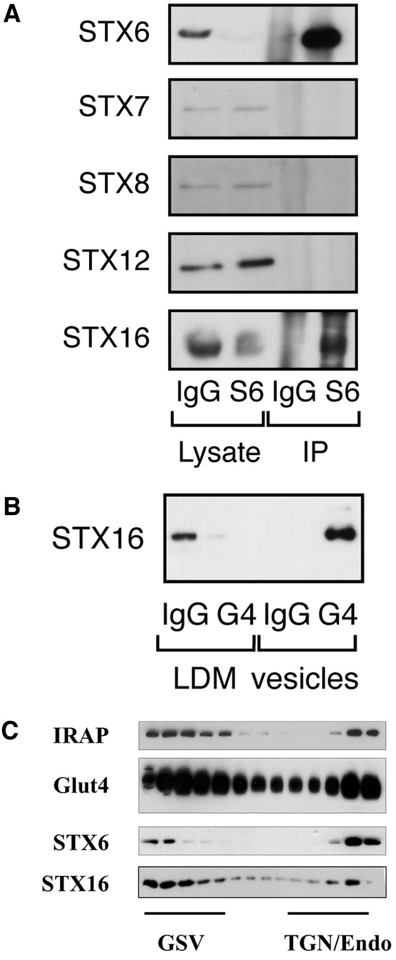
STX6 binds STX16. (A) Syn 6 was immunoprecipitated from lysates of 3T3-L1 adipocytes, as described in text. Anti-syntaxin 6 (mouse monoclonal) and anti-mouse Ig were used at 7.5 μg for cell lysates of a 10-cm dish. The representative immunoblot shown compares the lysate after IP with STX6 or IgG, and the corresponding immunoprecipitated material. In this figure, the lysate corresponds to 2.5% of a 10-cm plate of cells, and the IP corresponds to 15% of the total immunoprecipitate (i.e., 15% of the IP from a single 10-cm plate). (B) Presence within Glut4 vesicles of STX16 (see legend to Figure 2). (C) Iodixanol gradient profile of LDM fractions from basal (nonstimulated) adipocytes immunoblotted for Glut4, STX6, and STX16. The position of the GSV and TGN/endosomal peaks is shown. The representative experiment shown was repeated twice with qualitatively similar data.