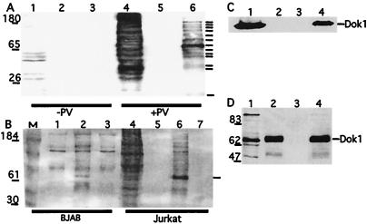
Figure 1.
Multiple p-Y proteins including Dok 1 bind specifically to wild-type SH2D1A in vitro. (A) Lysates from Jurkat T cells treated or not treated with pervanadate were incubated with GST (lanes 2 and 5) or with GST-SH2D1A fusion protein (lanes 3 and 6) bound to glutathione-Sepharose. Bound proteins were subjected to Western blot analysis with a p-Y-specific antibody. Arrows indicate major p-Y-containing proteins. The cytoplasmic cell lysates (1%) are in lanes 1 and 4. (B) Lysates from BJAB B and Jurkat T cells treated with pervanadate were incubated with glutathione-Sepharose-bound GST (lanes 1 and 5), -GST-SH2D1A (lanes 2 and 6), or -GST-SH2D1A-R32T (lanes 3 and 7). Lane 4 is 1% of a cytoplasmic lysate. Protein complexes were separated by SDS/PAGE and subjected to Western blot analysis with a p-Y-specific antibody. The position of a protein of ≈65 kDa precipitated by GST-SH2D1A is indicated by an arrow. All GST fusion proteins were expressed equally (data not shown). (C) Lysates (1% in lane 1) from pervanadate-treated BJAB cells were incubated with glutathione-Sepharose-bound GST (lane 2), -GST-SH2D1A (lane 4), or -GST-SH2D1AR32T (lane 3), and bound proteins were subjected to Western blot analysis with a Dok1-specific antibody. (D) Lysates (1% in lane 1) from pervanadate-treated BJAB cells were adsorbed onto GST-SH2D1A (lane 2), GST-SH2D1AR32T (lane 3), or p-Y antibody (lane 4). After gel electrophoresis and protein transfer, the membrane was probed with antibody to Dok1.

