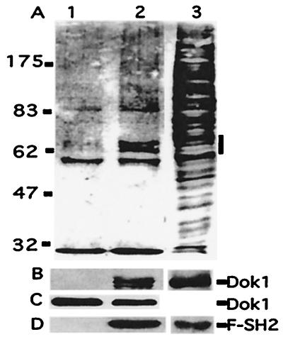
Figure 2.
Dok1 associates with SH2D1A in vivo. Lysates from BJAB or FLAG-SH2D1A1-converted BJAB cells treated with pervanadate were incubated with M2-FLAG-antibody beads (A, B, and D) or p-Y-specific antibody complexed to protein G-Sepharose (C), and immune-precipitated proteins (BJAB in lane 1 and BJAB/F-SH2D1A in lane 2) were subjected to Western blot analysis with p-Y (A), Dok1 (B and C), or FLAG (D) specific antibodies. The position of p-Y proteins migrating as a doublet and associated with F-SH2D1A is indicated by a small vertical bar to the right of lane 3 in A. The positions of Dok1 (B and C) and F-SH2D1A (D) also are shown. Lane 3 is 1% of the FLAG-SH2D1A-expressing BJAB cell lysate.

