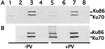
Figure 4.
Ku70 and Ku86 interact with SH2D1A in vitro. Cell extracts from Jurkat (A) or BJAB (B) cells treated (+PV) or without (−PV) pervanadate were incubated with bacterially expressed GST (lanes 2 and 6), GST-SH2D1A (GST-SH2) (lanes 3 and 7), or GST-SH2D1A-R32T (GST-SH2T) (lanes 4 and 8) adsorbed on glutathione beads, and the precipitates were analyzed by SDS/PAGE and Western blotting analyses. Lanes 1 and 5 are 1% total lysate. Filters were probed with a mixture of goat anti-Ku70 and anti-Ku86. The positions of Ku70 and Ku86 are indicated.

