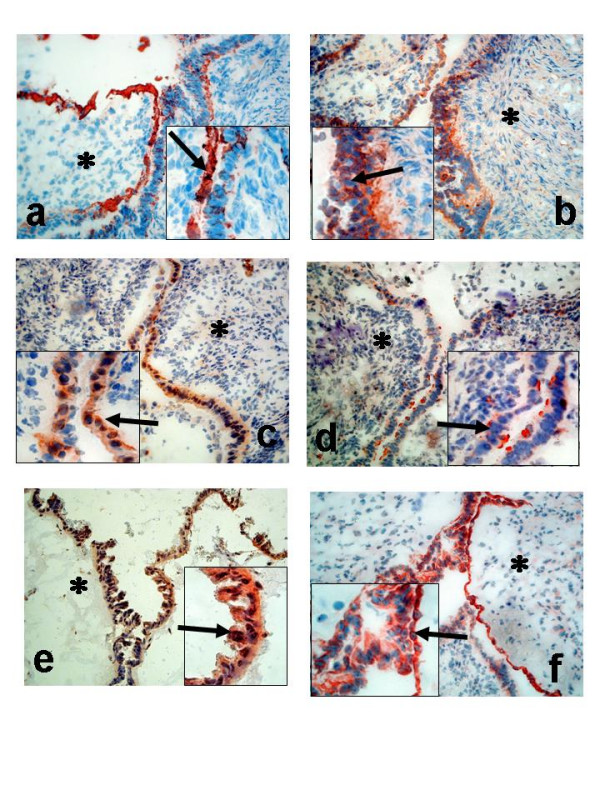
Figure 10.

Immunohistochemical analysis of ovarian tumours; Serous borderline type tumour, stage I (a-f); Staining with antibodies against CK8 (a); COX-1 (b); COX-2 (c); mPGES-1 (d);EP1 (e); EP2 (f). Arrow = epithelial cells, Star = stroma cells. (Original magnification ×200, inserted pictures ×400).
